
Various clinics as well as medical and sports centres took part in this study. Here, we are presenting charts which show the distribution of the index/coefficient of energy metabolism in certain organs and systems of organs. Values which correspond to the statistical norm are highlighted in green and have the following boundaries – from -0.6 to +1. Values outside of the normal range are less than -0.6 (red colour) and bigger than +1 (yellow colour).
A 56-year old woman came for an examination. She complained of having had nasal congestion and sore throat for approximately two months along with swelling of her legs in the ankle area and feet. From her medical history, we know that she has suffered from hypertension for long time, and started taking a special medicine for it – a calcium channel blocker – a month ago. She has had repetitive ARDs (acute respiratory disease) and cured sinusitis using conservative therapy. The patient has experienced occasional blunt chest pains during physical exercises, and has used Nitro Spray for pain relief. Objective examination has shown the following: the skin surface is pink and dry. The mucous membrane of the throat is hyperemic, but moist and clear. The auscultation of the heart showed clear sounds, vesicular breathing for lungs, and sno wheezing. Abdomen is not tense and palpation was pain free. The body temperature was 36.6 C. BP 135/90 mm. Pulse – 78 beats per minute.
The following results have been obtained using the crownscopy technique: the level of energy metabolism in the maxillary area, frontal and paranasal sinuses and the area of the ethmoid bone are below the threshold. Significantly lower energy values appear in the ankle and feet area. The sector which is responsible for the coronary arteries is also in the area of energy deficit.
The aggregate set of complaints, medical history and the results of the examination indicate a chronic inflammation in the paranasal sinuses, the edema of the lower part of the legs as a side effect of the taken medicine (this is listed in the side effects for the medicine), as well as angina and arterial hypertension. These clinical observations are reflected in the changes of the crownscopy results which are shown in the figures further:
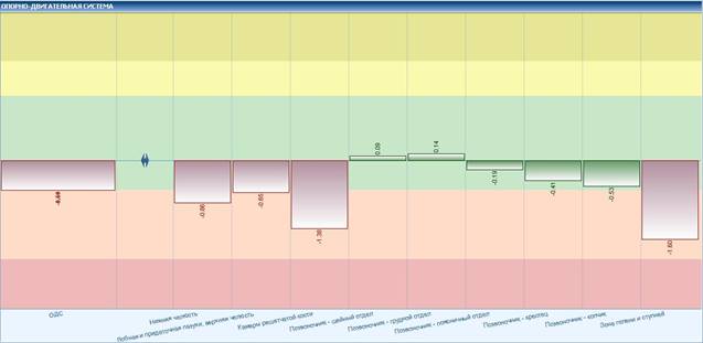
Fig.1 Musculoskeletal system. Hypofunction in the area of the paranasal sinuses and lower part of the legs.
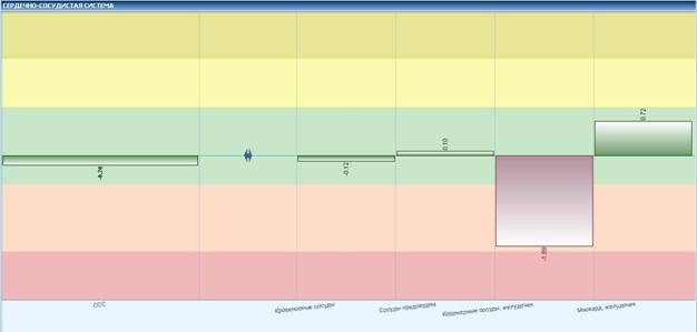
Fig.2 Cardiovascular system. Hypofunction in blood supply by coronary arteries/artery.
A 28-year old female complains of having had weakness, fatigue (mostly towards evenings), sleepiness, and depressed mood for approximately 20 days. Her body weight tends to increase (+1200 g within 2 months). It is known that the patient has suffered the aggravation of the chronic pyelonephritis twice within the last 9 years and had to stay in a hospital. The study of the crowngrams shows a significant decrease in the functional activity of the thyroid gland. Also, we should note the energy deficit in the segment which is responsible for the uterus and pancreas functions (a very slight deviation from the norm). From the conversation with the patient, we get to know that she has been experiencing menstrual disorder in the past 1.5 years. The cycle could grow to 38-45 weeks, and then change to oligomenorrhea. The patient has not obtained medical advice on this issue. The patient was recommended to visit a gynecologist for an in-depth diagnosis of the menstrual disorder, as well as taking clinical and biochemical blood tests, a test for thyroid hormone levels with a consequent visit to an endocrinologist. The medical history and the results of crownscopy show insufficient functional activity of the above mentioned areas and allow excluding endocrine pathologies.
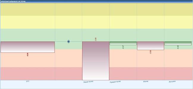
Fig.3.Urogenital system. Hypofunction of the left kidney and the uterus.
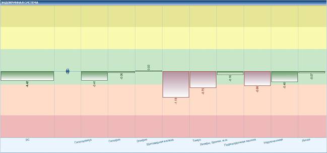
Fig.4. Endocrine system. Energy deficit in the following areas: thyroid, pancreas, and thymus.
A 51-year old male complains of having pains in the lumbar area of the spine which extend into the left thigh. We know that he has spent the most part of the last three days in a sitting position (about 15 hours of driving). The crowngrams show the energy deficit in the lumbosacral and the coccygeal areas of the spine. However, no other areas with significant decline in the energy metabolism have been identified. The results correspond to the assumption of the changed blood supply in the region due to the forced posture.
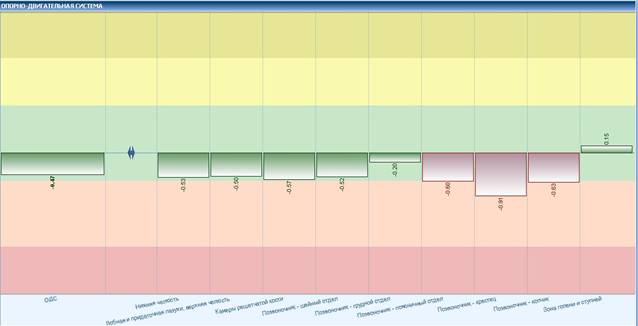
Fig.5. Musculoskeletal system. Energy deficit in the lumbar area extended to the tailbone.
A 24-year old male felt acutely ill seven days ago with the appearance of muco-purulent discharge coming from his nostrils, cough and difficulty with excreting the phlegm, along with an increased body temperature of 37.8 C and muscle-joint pains. He has been treated with expectorants, antihistamines, and non-steroidal anti-inflammatory medicines. He noted significant improvement in the condition by the time of the examination. He still had running nose and occasional cough with clear phlegm, while the body temperature reduced to 36.6 C. The lung examination showed vesicular breathing, isolated rales in the lower part of the lungs. The analysis of the crowngrams shows a decline in the energy performance of the upper respiratory tract which corresponds to the decay of the exacerbation of the chronic inflammatory process. The reduced energy levels in the respiratory and lymphatic systems as well as an unsatisfactory value of the adaptation index indicate the chronic inflammation.
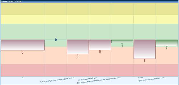
Fig.6. Respiratory system. Energy deficit of the sinuses and the lower respiratory tract.
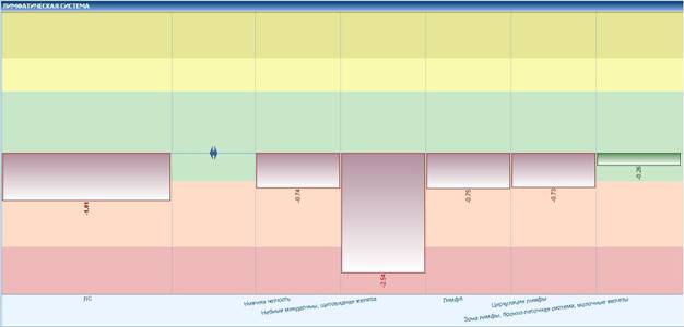
Fig.7. Lymphatic system. Hypofunction of the lymphatic system.
Crownscopy in sports medicine: opportunities and use cases
Sports medicine sees its main goal as medical and biological preparation of athletes via the resolution of several tasks. Modern crownscopy methods enter the scope of practical approaches of sports clubs and centres along with traditional methods for control and diagnosis.
The selection of the kind of sport is one of the first tasks when working with a young athlete. The application of the crownscopy technique at this step helps to identify the health compensation levels at the moment of the examination, as well as determine the level of physiological resources of the athlete.
Let us have a closer look at the initial evaluation of the athlete’s condition. The diagram of the energy metabolism in the tissues shows that the production and consumption of the ATF in every system is in dynamic balance (for example, the average value for the digestive system is +0.4, which corresponds to the normal level of energy metabolism). The adaptation values show that the functional reserves of the examined athlete are within the sufficient functional range. Based on the obtained data we can assume that the person is healthy and his inner reserve is on a high level which allows getting even fitter and achieving good results.
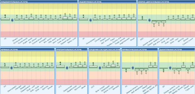
Fig.1 Energy distribution in the organs and systems. In general, every system and individual organs show normal energy levels.
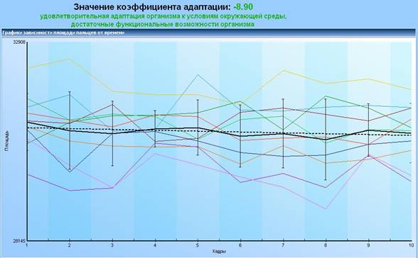
Fig.2. Calculations of the adaptation coefficient
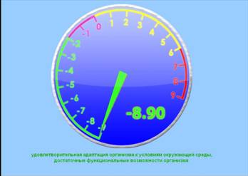
Fig.3. Adaptation coefficient within the normal range.
Adaptation is a set of reactions and mechanisms which provide for the functionality of the organism in various environments. The crownscopy method allows calculating the degree of adaptation as a numerical coefficient, which is based on dynamic characteristics of the inner body reserves.
Any energy deficient zone draws attention of a doctor or a trainer to the fact that the patient may either have physiological limitations at the moment or they might occur due to high and unusual loads.
For example, let us have a look at the following chart.
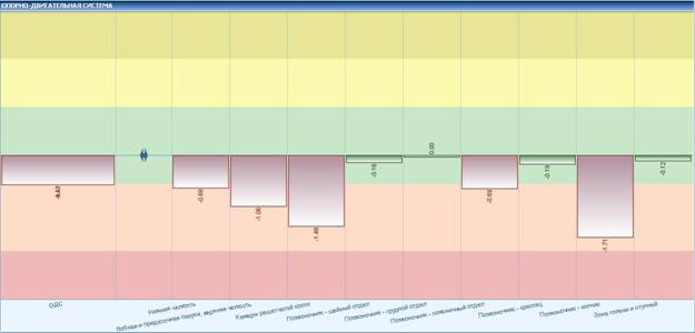
Fig.4. Energy distribution in the musculoskeletal system.
We can see that despite the patient has no complaint at the moment the deficiency zones are still present. The patient admits that he has frequent colds and hurt his back several years ago hence now he experiences occasional pains in the lumbar region. These changes are reflected in the crowngrams and interpretation charts. Such factors become prerequisites of more difficult conditions which are not ideal either for sportsmen or for trainers.
Crownscopy allows monitoring the functional state of an athlete during an individual training or after a course of trainings taking into account additional conditions (for example, extra sports equipment, harder workouts, and protein drinks).
For example, evaluation of the energy condition of an athlete in the beginning of the training (volleyball game) and 40 minutes after the start of the game show the decrease in the energy levels in the musculoskeletal system.
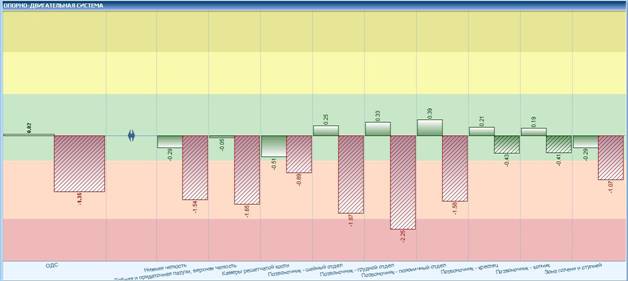
Fig.5 Energy distribution in the musculoskeletal system. Red lined bars indicate energy deficit which appeared 40 minutes after the start of the training. Green bars show initial values.
Musculoskeletal system. Lined bars show energy levels 40 minutes after the beginning of the training.
Thirty minutes after the end of the training the energy distribution chart shows the growth of the energy balance beyond the original values. This indicates satisfactory fitness and rapid restoration of the functional reserves.
Thus we can assess the fitness change by measuring the initial energy indexes and the same indexes after the end of the physical activity.
Mental state of an athlete is one of the main keys to success. Good mental health provides will to overcome difficulties and allows for effective competition. Evaluation of the patient’s mental state is one of the applications of crownscopy. The dynamics of this index achieved as a result of a series of consequent measurements may indicate an unstable psychoemotional state of the athlete.
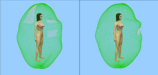
Fig.6 Psycho energy field. An example of the change in psychophysiological state of the patient before and after rest and a motivational conversation with the trainer. The right image shows the improved state. Graphically, the improvement is shown with the green shape getting a smoother oval form and the disappearance of the red zones.
Crownscopy allows growing one’s fitness capacity via timely control over the functional state of the organism, which does not take long to implement and does not require time-consuming analysis of the results. Methods of biological feedback (BF) are used as well.
BF – biological feedback – is implemented as a computer program where a user solves tasks by activating psychological capabilities (for example, by visualizing the start of the swimming competition or snapping of the ball in a game, or a win in the competition) and alternating it with relaxation (visualization of one’s rest). Managing their condition, the athletes will grow stress resistance and the ability to change their state depending on the situation (for example, a quick switch from the maximum stress during a competition which causes overstimulation of the nervous system to a calm state).
Thus crownscopy may provide invaluable help in medical and functional monitoring in sports, help to improve athletic performance and facilitate recovery.
For Healers
The method of crownscopy can be used by a wide range of specialists in both traditional and alternative medicine in everyday practice and while conducting complex scientific researches.
For example, comparison of the change in the patient’s states before and after a corrective procedure.
We are going to discuss the case in which one session of craniosacral therapy with the duration of 40 minutes has been offered to the patient. We can notice the growth of the following values when comparing the indexes: the area of the crown luminescence has grown from 21382 to 25511, the normalised area has grown from 0.46 to 0.55, and the symmetry shows an increase by 0.06%. Visually, it is possible to register the expansion of the energy field with the closure of several vacant states.
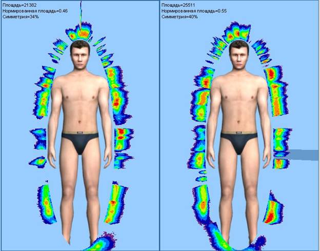
Fig.1. Energy state of the patient. When comparing images before and after we see the growth of the luminescence area (from 21382 to 25511), the growth of the normalised area and symmetry.
We can also note the positive changes in the psychoemotional state:
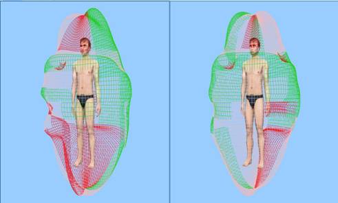
Fig.2. Psychoenergy state. Gradual formation of a more rounded shape of the psychoenergy field with the completion of the energy deficit up to the norm.
We can conclude that the chosen method has had positive impact on the patient.
Another example – the Reiki techniques. The measurement has been taken three times within an hour. 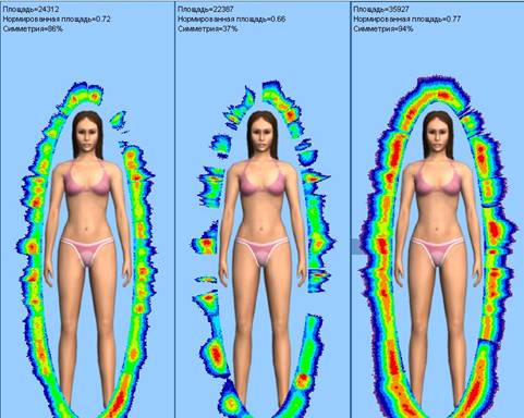
Fig.3. Energy state. Triple measurement. Visual proof of the energy field growth.
We would like to note very interesting dynamics. At first, the values of all indexes would drop, and then considerable growth in the energy metabolism levels in all organs and systems.
This is what it will look like on a diagram of distribution over organs and systems:
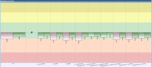
Fig.4. Energy distribution in the nervous system before and after the treatment. Lined bars show energy levels after the treatment.
The obtained crownscopy results may be used when working with the client (for example, visualization of certain changes in the parameters which are being corrected), may be saved into an archive, and may be used for visual assessment the state before and after the treatment.
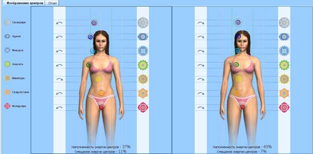
Fig.5.Energy centres shift closer to the median line after the correction, with their fullness growing.